
Professor of Materials Science
BSc University of Bristol
MSc University of Bristol
PhD University of Bristol
Electron Microscopy
My current research is based on the development and application of new electron microscopy techniques to study the structural and functional properties of a variety of materials with high spatial resolution in two and three dimensions.
Electron Tomography
By recording a tilt series of transmission electron micrographs, using a variety of imaging and analytical modes, electron tomographic techniques are being developed to provide high fidelity 3D reconstructions, with ca. 1nm3 resolution, of morphology, defect structure, composition and electrostatic potentials across a range of materials and devices. Implementation of novel reconstruction algorithms, including compressed sensing to incorporate a priori information, yields high quality reconstructions from relatively few images and provides a platform for atomic resolution reconstructions. We have also a research program developing ‘mesoscale’ tomography in which use of a dual beam SEM-FIB, coupled with EDX and EBSD techniques, provides 3D reconstructions of morphology, crystallography and composition, with spatial resolution and fields of view that bridge the gap between (S)TEM-based and X-ray tomography.
Nano-Plasmonics
Using monochromated electron beams, STEM EELS maps (in both 2D and 3D) from plasmonic structures are used to determine fundamental optical excitations of metallic and composite nanostructures. By using blind source separation techniques, in conjunction with more conventional spectral and image processing methods, high quality maps of localized surface plasmon resonances can be determined. The dispersion of low loss excitations is also being studied including the use of hybrid ω-q modes. Complementary theory, and associated simulations, is being undertaken to understand the complex low loss spectra from nanostructures and to correlate with near field excitations and optical spectra.
Structure Determination using Electron Diffraction
Precession electron diffraction (PED) has become a cornerstone technique for many electron crystallographers studying inorganic structures. We continue to develop PED (both in zone axis mode and as a tilt series), combined with novel structure solution and refinement algorithms, to determine the crystal structures of many previously unknown phases (e.g. multi-phase oxides, pharmaceuticals, MOFs). A scanning variant (SPED) may be used to determine orientation relationships with nm spatial resolution and by combining this with tomography, full 3D orientation mapping of nanoscale structures may be found. Structured diffuse scattering is being analysed quantitatively to determine thermal motion and phonon dispersion in important oxide and semiconducting materials.
Nanoscale Structures
Semiconducting nanotubes and nanowires, catalysts, filled elastomeric materials and metallic nanoparticles are being studied by high resolution (aberration-corrected) imaging, analytical electron microscopy and electron tomography to correlate their atomic and nanoscale structure to underlying physico-chemical properties.
|
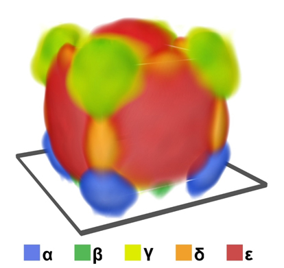
Composite colour image showing five surface plasmon modes (labelled α - ε) on a silver nanocube (100nm across) reconstructed using 4D spectrum-tomography (see O. Nicoletti et al., Nature, 502 80-84 (2013).)
|
 |
- Midgley P.A. and Dunin-Borkowski R.E., "Electron tomography and holography in materials science", Nature Material,s 8 271-280 (2009).
- Saghi Z., Holland D., Leary R.K., Falqui A., Bertoni G., Sederman A.J., Gladden L. and Midgley P.A., "3D Morphology of Iron Oxide Nanoparticles with Reactive Concave Surfaces – a Compressed Sensing-Electron Tomography (CS-ET) Approach", Nano Lett., 11 4666-4673 (2011).
- Eggeman A.S., Illig S., Troisi S., Sirringhaus H. and Midgley P.A., "Measurement of molecular motion in organic semiconductors by thermal diffuse electron scattering’ Nature Materials", 12 1045–1049 (2013).
- Nicoletti O., de la Pena F., Leary R.K, Holland D., Ducati C. and Midgley P.A., "Three-Dimensional Imaging of Localized Surface Plasmon Resonances of Metal Nanoparticles", Nature, 502 80-84 (2013).