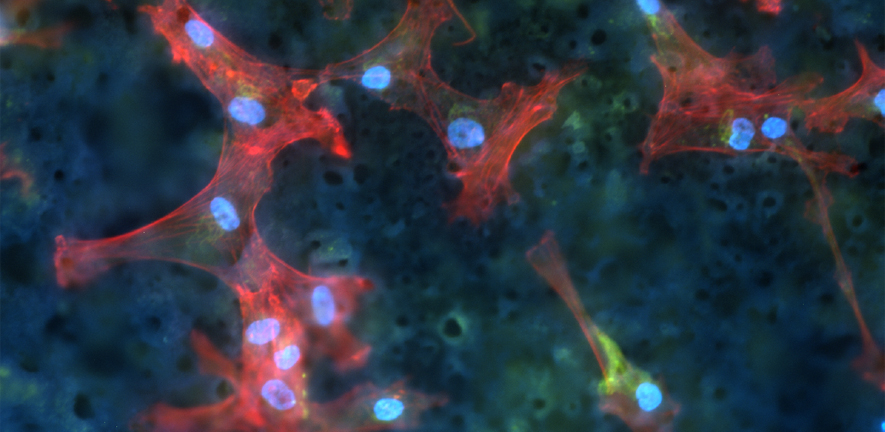
Banner image: Actin (red), nuclei (blue) plasma membrane (green) immunofluorescent images (10 x) of osteoblast cells cultured for 18h on β-Ti45Nb. (Image provided by Dr Edi Tanase)
2D and 3D imaging techniques are central to research in Materials Science. We have state of the art 3D imaging facilities ranging from high tilt-range electron microscopes through X-ray microtomography to confocal laser scanning microscopy which allow comprehensive 3D imaging of structures at the length-scales from nanometres up to millimetres. Tomography based approaches typically combine a large number of different projections of an object to reconstruct a representative volume using computer algorithms. Confocal approaches allow a series of images at different depths through a sample to be recorded and again combined to produce the original volume.
The applications of such imaging techniques are numerous, from understanding how the shape and surface chemistry of catalysts affects their ability to affect chemical processes through to studying medical implants and structures containing biological cells.
The 2D images recorded using microscopy approaches can also provide valuable information about the samples, for example the image above shows how fluorescence microscopy allows labelling of specific features in biological samples. Other approaches include chemical and structural analysis through scanning transmission electron microscopy combined with statistical decomposition to isolate different components in microstructures.